This site
is under construction. However, here is a sample of what
we hope to provide in the future.
Click on thumbnails to see a full-size
version
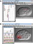
This
panel shows the various muscle cells within a nematode and
the structure of sarcomeres in C.elegans muscle.
This
image shows the lineage derivation of all the body wall muscle
cells. The picture is modified from Sulston et al, 1983 Dev.
Biol. 100:64-119.
This
picture is from Schnabel et al 1997 Dev. Biol. 184:234-265.
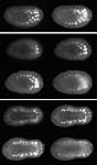
These
panels are unpublished images of myoD expression through
development.
Early development
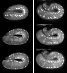
MyoD
expression - intermediate development
MyoD
expression - comma stage
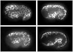
MyoD
expression - two-fold stage
This
picture is from Moerman et al 1996 Dev. Biol.173:228. It shows
the migration of muscle cells from their lateral position in
the embryo to their final positions where they will form the
dorsal and ventral muscle quadrants on the left side of the
animal.