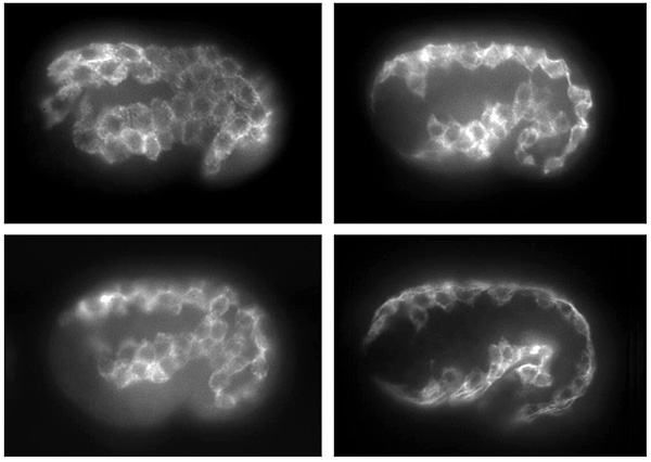
These four panels show an embryo from comma
to almost the two-fold stage of development. Anterior is to the
left and dorsal is towards the top in all four panels. This figure
illustrates how the muscle cells are intially positioned on the
lateral side of the animal and form a sheet of muscle cells.
This figure shows only the left lateral side. Over time this
sheet separates starting at the anterior end and two rows of
cells move to the ventral lateral surface and two rows move to
the dorsal lateral surface. Eventually there will be four quadrants
of muscle cells (two quadrants arising from each side). In the
earliest image the signal from the thick filamant protein can
be detected throughout the cytoplasm but later it is more restricted
and the cells have become more polarized. This restriction represents
the formation of the muscle sarcomeres adjacent to the plasma
membrane which is in intimate contact with the basement membrane
and the underlying hypodermis.