LAB 8 : CIRCULATORY AND RESPIRATORY SYSTEMS

Objectives:
1)Understand the process of respiration and circulation in each animal and be able to state the similarities and differences between them.
2)Learn the anatomy of the heart and the blood vessels branching to and from it in each animal.
3)In the dogfish, understand how blood flows from the heart and through the gills to allow for gas exchange.
4)In Necturus, understand how blood circulates through the heart to the gills and lungs.
5)Use the rat/cat to learn the names of the membranes that line the lungs, heart and body cavities.
6)In the mammal, understand the process of respiration
7)In the mammal, understand how blood flows through the heart, to the lungs, and to the rest of the body.
8)Learn how the heart and aortic arches have evolved.
DOGFISH RESPIRATORY SYSTEM
In the shark, the circulatory and respiratory systems function as one because the heart pumps unoxygenated blood returning from the body to the gills for oxygenation. From the gills, the oxygenated blood is distributed to the body. Gas exchange also takes place in the skin, but primarily in the gills.
Look at the diagrams and use your specimen to follow the passage of water during respiration. Water enters through the mouth and spiracles (which have a one way valve) and exit through the five gill slits. To draw water into the mouth, the gill slits close and the pharyngeal chamber expands to suck in water. When the pharynx is filled, the mouth closes and the gill chambers expand and fill with water. Then the gill slits open and the chambers constrict to flush out the water.
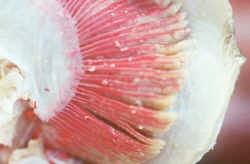
If you look down the gill slits, on each side you will see one half of the gill, a demibranch. The internal septum, blood vessels, nerves, muscles and two demibranchs make up each holobranch. At their base are gill rakers, which project into the mouth and protect the gills from mechanical injury. The demibranchs have primary lamellae, which can be easily seen, and secondary lamellae, which can be seen if you remove a portion of the gill and examine it closely. The oxygen rich water flows in a countercurrent pattern to the blood and allows efficient oxygen and carbon dioxide exchange to take place.
Dogfish Heart and Aortic Arches
To expose the blood vessels, first skin the roof of the mouth where the efferent (e=from) branchial arteries are located. They are pink and usually well injected. Make a shallow cut just inside the teeth and peel the skin off by flaying it. You will have to go well down the pharynx to the place where the arteries meet.
The floor of the mouth, under the attached tongue, has the afferent (a=to) branchial arteries and posteriorly the heart. The yellow or blue latex does not make it through the heart so the vessels are not injected and are delicate. They are colourless or brown with blood. Remove only the skin with a scalpel. Expose the heart by removing its protective cartilage by nicking the posterior sides and pulling it forward. Identify the sinus venosus, auricle and ventricle. Trace the ventricle anteriorly into the conus arteriosus, and into the short ventral aorta.
Using your diagrams, trace the pathway of the blood through the afferent branchial arteries, the gills and the efferent branchial arteries.
Pry off the cartilage on the afferent vessels with forceps and tease it loose with dissecting needles but do not use the scalpel any more in this area. The vessels are almost stuck to the cartilage. Expose all five afferent branchial arteries as far as the right gills. Follow one into the gill as far as you can. Expose the efferent arteries on each side of this afferent one and find how the major efferent duct loops up to the proceeding gill. In the region of the gills, skin a pair of gill arches and trace one loop of the efferent branchial arteries around a gill slit.
Learn how the gills work by answering the following questions:
*How many afferent arteries are there and what direction does the blood flow in them?
*How many efferent arteries are there and what direction does the blood flow in them?
*Where does the blood become oxygenated?
*Where does the unoxygenated blood enter the gills?
*How does it become oxygenated?
*What is the path of water flow over the gills?
*How does oxygenated blood leave the gills?
*How are the afferent and efferent blood vessels of the gills connected to each other within the gills? Describe in your own words.
*Describe the efferent loop around a gill slit.
*Describe the path of the blood through the heart.
NECTURUS RESPIRATORY SYSTEM
Much of the respiration in amphibians is through the skin. Up to 75% of the oxygen and 90% of the carbon dioxide is exchanged this way in aquatic forms. In addition, Necturus has three pairs of gills and one pair of lungs. External gills develop before the two gill slits open and can be waved about by branchial muscles. Lungs developed from swim bladders, which sometimes function as lungs in fish that gulp air. Look for the glottis, a longitudinal slit in the pharynx floor, which is the opening to the lungs. The larynx is a pair of cartilages surrounding the glottis and will not be noticed. The trachea is short. The lungs are long, slender and saclike. They run along the dorsal sides of the pleuroperitoneal cavity and are attached to the body wall by the pulmonary ligament on the left side and on the right, the hepatocavopulmonary ligament that also supports the liver. The lungs are used as hydrostatic organs and only contribute about 2% of the gas exchange. Air is gulped via the mouth as it is in lungfish.
Necturus Heart and Aortic Arches
Carefully skin the insides of the pharynx, top and bottom, concentrating on the area between the gills to the transverse septa. Expose the heart on the ventral floor of the mouth and the afferent arteries leading to the gills. Use a dissecting microscope if necessary.
The heart should be viewed from the mouth. It consists of a left and right atrium, which appear as one dorsally but wrap on either side of the bulbous arteriosis. The single, large, muscular ventricle is ventral, usually blue, and posterior to the atria. The sinus venosus is blue, dorsal, and posterior to the atria. Blood enters the sinus venosus via the lateral common cardinal veins and the posterior, postcaval vein. Unoxygenated blood enters the heart from the body via the sinus venosus to the right atrium and oxygenated blood from the lungs enters the heart via the left atrium. It enters the ventricle in separate pulsations. The ventricle pumps into the bulbus arteriosus.
Follow the branches of the ventral aorta and its branches to the gills (afferent arteries) and head (external carotid artery). On the roof of the mouth, find the efferent arteries from the gills, internal carotid going to the head and subclavians going to the arms. Note the thin pulmonary artery, which leaves the junction of the two posterior most efferent arteries. These vessels join the dorsal aorta, which carries oxygenated blood to the body.
QUESTIONS:
*How many afferent arteries are there and what direction does the blood flow in them?
*How many efferent arteries are there and what direction does the blood flow in them?
*Where does the blood become oxygenated?
*Describe the path of the blood through the heart.
MAMMALIAN RESPIRATORY SYSTEM
Open the thoracic cavity by cutting through the diaphragm, which separates the abdominal from the thoracic cavity. Cut through the ribs on the left side of the sternum, leaving the sternum in position. Lift the ribs to look sideways at the thoracic cavity. The object here is to see the mid ventral membranes before removing the sternum.
Note the shiny membranes covering the lungs and the inside of the thoracic cavity. These are the visceral and parietal pleura. Note also the mediastinum which is the shiny membrane dividing the right and left pleural cavities.
Show others in your group the mediastinum and then remove the sternum by cutting through the mediastinum and anteriorly to expose the trachea. Be careful not to damage the heart or the accompanying blood vessels.
The heart lies in the center of the thoracic cavity at the posterior end. It has a thin, transparent parietal pericardium around it.
The lungs are the brownish lobes to the right and left of the heart. Entering the lungs on either side from the trachea are the bronchi. They are supported, as is the trachea, by incomplete cartilage rings.
So that you will not destroy the heart, use the displays of injected lungs to see the divisions and subdivisions of the bronchi.
They divide into: secondary bronchi, one to each lobe and bronchioles, within each lobe. Too small to see are the small end branches, the infundibula, which are lined with alveolar sacs. Different animals vary in the number of lobes to each lung. The cat and rat have four right lobes, humans have 3. The cat has 3 left lobes, rats have one and humans have two.
In your specimen, trace the trachea anteriorly. The rings supporting it are incomplete. Note that the esophagus lies dorsal to the trachea and the gaps in the rings allow the esophagus to attach on the dorsal surface of the trachea. Trace the trachea anteriorly to the larynx, or voice box. Note that it is supported by cartilages. These are the hyoid bone and thyroid and cricoid cartilages.
What is the evolutionary origin of these cartilages?
The air enters the nasal passageway at the nostrils and is separated from the oral passageway by the hard and soft palate. A nasal septum divides the air from the two nostrils. You should be able to look down the pharynx and see a triangular flap, the epiglottis which moves up to open the air passageway (glottis) or down to close the air passageway and allow food down the esophagus. Inside the larynx at the base of the epiglottis are two lateral flaps, the vocal cords, which open and close to produce sound.
In the upper part of the pharynx, slit the soft palate to see the nasal passage. Into the upper back of this passageway opens the eustachian tubes which lead to the middle ear. This opening allows you to "pop" your ears, thus equalizing the pressure on both sides of the tympanic membrane to prevent it from rupturing.
Mammalian heart and aortic arches
Remove the thymus gland on the anterior of the heart. It is absent in older animals. The heart lies in the pericardial cavity and is enclosed by the pericardial sac. In the cat, this sac has to be removed. The heart consists of two atria, and two ventricles. The sinus venosus seen in the dogfish and Necturus is now incorporated in the wall of the right atrium and is now called the sino-atrial (SA) node. This is the ‘pacemaker’ of the heart, setting the rate of muscular contraction (heart rate). The auricles, or atria, are small dark structures on the antero-lateral border of the heart. They are divided from the ventricles by a groove, and have much thinner walls than the ventricles. The ventricles appear as one externally, but if you feel them, the larger left ventricle is hard due to its thick walls, the smaller right ventricle is soft as its walls are thinner.
When you have finished finding the arteries and veins below, slit open the ventricles from the posterior tip towards the atria, into top and bottom pieces, and lift up the top flap to see the division of the ventricles. The valves are small and best observed in the models and plastic pig heart.
Deoxygenated blood enters the heart from the body via the branched superior vena cava (precava), which drains the head and arms, and the inferior vena cava (postcava), which drains the abdominal cavity. These both enter the right atrium and are injected blue. In the rat, the left branch of the superior vena cava swings to the left and under the heart to join the inferior vena cava. In cats and humans they join anterior to the heart.
From the right atrium, deoxygenated blood flows through a tricuspid valve into the right ventricle which pumps the blood through a semilunar valve to the blue injected pulmonary aorta (VI) which divides into two pulmonary arteries, one to each lung. In the lung the blood vessels subdivide into capillary beds around the alveoli, where gas exchange takes place.
The oxygenated blood flows back from the lungs via the colourless pulmonary veins, which run a parallel, course to the pulmonary arteries but enter the left atrium. From the left atrium the oxygenated blood flows through the bicuspid (mitral) valve to the left ventricle.
The muscular left ventricle must pump oxygenated blood to the entire body including the heart itself. Blood is pumped through the semilunar valve to the systemic aorta (IV). The coronary arteries branch off of the aorta to supply the surface of the heart. The systemic aorta then bends to the left as the aortic arch. At the top of the arch, vessels branch off to supply the arms and head. In the rat and human there are three main vessels branching off the aorta, first the brachiocephalic, or innominate, (supplying the right arm and head), then the left common carotid (III; to the head) then the left subclavian (IV; to the left arm). In the cat the left common carotid branches off the brachiocephalic rather than having a separate origin and therefore there are only two branches off the aorta. The brachiocephalic sends a branch, the right subclavian (IV) to the right arm and continues as the right common carotid (III) to the head. The common carotids travel up the neck on either side of the larynx and supply the thyroid, larynx, throat, and face muscles, tongue and part of the brain. The internal carotids supply the deep parts of the head; the external carotids supply the more superficial areas.
After the head and arm vessels leave the aortic arch the systemic aorta continues towards the diaphragm as the thoracic aorta and posterior to the diaphragm as the abdominal aorta. These are equivalent to the dorsal aorta in the dogfish.
Comparative Aspects
The purpose of this section is to give you an understanding of:
A)The increase in complexity that has occurred from the two-chambered fish heart to the four-chambered mammal heart. To study these features examine the models of hearts and aortic arches displayed in the lab, the illustrations in your lab and textbook and what you have learned about the dogfish, mudpuppy, cat and rat.
B)The loss in number of aortic arches in the evolution from fish to mammal, and the change in function of those arches that remain.