THE DIGESTIVE AND UROGENITAL SYSTEMS

Objectives:
1)Learn the names of the body cavities and the membranes that line them.
2)For each animal, identify and learn all of the digestive organs and features of the digestive tract.
3)Understand the function of each digestive organ.
4)Use the rat to learn glands in the neck.
5)For each animal, identify and learn all of the structures associated with the reproductive and urinary tract.
6)Understand the process of gamete formation, fertilization, and birth in each animal.
7)Learn the glands associated with the reproductive system in the cat and rat.
8)Be able to compare and contrast each system in the three animals.
BODY CAVITIES
Vertebrates have a coelomic body cavity. This coelomic space is divided anteriorly into a pericardial (heart) cavity and a posterior pleuroperitoneal cavity by the transverse septum, a tough, white membrane. This is the situation in the dogfish and Necturus. In mammals, a muscular diaphragm forms a separation between two large cavities. The anterior cavity is called the thoracic cavity and is subdivided into a central pericardial cavity surrounding the heart and paired lateral pleural cavities around the lungs. The esophagus runs through this cavity, but we will be looking primarily at the posterior cavity, the abdominal cavity in this lab.
The posterior pleuroperitoneal (abdominal cavity in mammals) cavity houses the liver, digestive tract, and gonads. This body cavity has muscular walls (mesoderm). The cavity is lined on the inside with a transparent parietal (somatic) peritoneum. It is attached to the muscles of the body wall but it also overlies the urogenital system, which is retroperitoneal. The parietal peritoneum from each side meets dorsally and ventrally to form a double walled mesentery. This splits to line the digestive tract and other organs and is called splanchnic (visceral) peritoneum. The peritoneum is serous (wet). The fluid serves as a lubricant to allow frictionless movement of the organs.
The sheets of membrane suspending viscera and joining them with each other are called mesenteries. The primary mesenteries are dorsal and ventral, although the dorsal mesentery is often interrupted and moved to one side with the organs and the ventral mesentery is reduced to the membranes of the liver and bladder. Mesenteries running from organ to organ are usually called ligaments.
DIGESTIVE ORGANS
The digestive tract is a tube, which begins at the mouth and ends either at a cloaca or anus. It processes food, which moves by peristalsis through the process of digestion, absorption and elimination. The general pattern is to have an oral (buccal) cavity, pharynx, esophagus, stomach and intestine. Accessory organs are the pancreas, liver and gallbladder, which arise as evaginations from the embryonic digestive tract. We will be looking at the variations and similarities in the digestive tracts of the dogfish, salamander and mammal in this lab.
THE UROGENITAL SYSTEM
The urogenital system is a combination of the two closely related systems – the excretory and the reproductive. The primitive kidney is built for osmoregulation and functions in marine, freshwater and land environments. Nitrogenous waste can be removed by diffusion through the skin in aquatic environments, but on land, the removal of nitrogenous waste is a primary function of kidneys. Kidneys consist of several renal corpuscles (glomerulus in a Bowman’s capsule) each connected to a tubule leading to a common tubule which is segmentally attached to a longitudinal collecting duct.
Gonads produce gametes and maintain secondary sex organs (reproductive ducts and glands) and secondary sex characteristics. They arise from genital ridges medial to the nephric tissue. Germ cells migrate into the genital ridges. Genes and hormones determine whether the germ cells will form ovaries or testes.
Eggs are released into the coelom before entering the oviduct. Sperm is always in a closed system of vessels. Fertilization may be external or internal. External fertilization is always oviparous in which eggs are laid, sometimes with yolk, and development takes place externally. If the eggs have a shell or capsule, fertilization must take place internally before the coating is place on the eggs. Live young are the result of internal fertilization. Ovoviviparous development takes place without direct nourishment from the mother, although yolk is usually present. In viviparous development, there is a placenta to nourish the embryo.
Once you have studied the reproductive structures of your animal, you must trade animals with a group that has an animal of the opposite sex. It is the therefore essential that you do not destroy any of the reproductive features.
DOGFISH
Dogfish digestive tract
Dissection: Use scissors to cut through the body wall, from the cloaca to the transverse septum, making sure your scissors do not cut underlying organs. Make your longitudinal cut off-center from the midventral line and out to the pectoral and pelvic fins, then turn back the flaps.
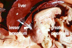
Note the falciform (=suspensatory) ligament, which is a remnant of the ventral mesentery, hanging midventrally from the liver in the anterior half of the pleuroperitoneal cavity. You will have to cut this ligament to look into the cavity so show the group first. Try to leave the ligaments and mesenteries intact. Other remnants of the ventral mesentery are the lesser omentum from the liver to the stomach and small intestine and reproductive mesenteries, which will be looked at later. The dorsal mesentery is seen as the greater omentum holding the esophagus and stomach to the dorsal body wall and dorsal mesentery holding the intestine. From the stomach to the spleen is a gastrosplenic ligament.
The oral (buccal) cavity contains the tongue and teeth. The pharynx is the portion leading past the spiracle and five gill slits and also contains the tongue in dogfish. Note the taste buds on the tongue.
To view this area, cut through the corner of the left jaw of the dogfish and perpendicularly through the center of the gills to the pectoral fin. Fold a paper towel over the teeth and use your scissors to open the mouth wide. Make sure you cut all the way through to the pharynx. Cut horizontally across to the other pectoral fin, making sure you are posterior to the transverse septum. You do not want to cut into the pericardial cavity. You will have to cut through the esophagus until you get to the gill slits on the other side. Pull the lower jaw open and clear out any debris.
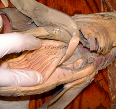
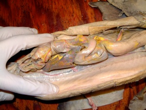
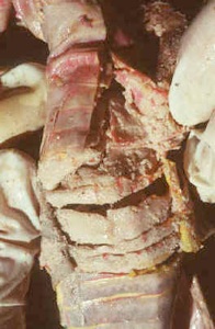
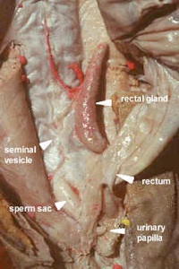
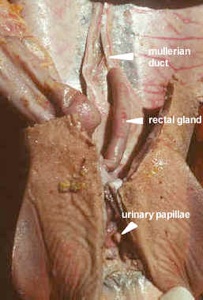
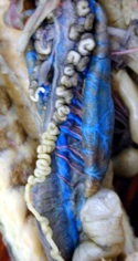
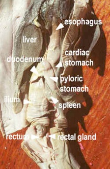
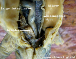
The esophagus extends from the pharynx at the transverse septum and is lined with papillae, stumpy finger-like projections, which form a tight seal to keep water out. Cut into the esophagus to see the papillae and the stomach with its longitudinal folds called rugae. The "U" shaped stomach, with an anterior cardiac limb and a posterior pyloric limb ends in a constricted pyloric sphincter. The anterior portion of the small intestine is the duodenum and the posterior portion if the ileum, which contains an internal subdivision, called the spiral valve. Cut into the ileum to see the valve, which increases the surface area of the intestine. The large intestine (rectum or colon) is a shorter section than the small intestine has a rectal gland entering it. The rectal gland is for salt excretion for osmoregulation. The rectum, the most posterior portion, ends in the anus which projects into the cloaca, a common opening with the urogenital ducts.
Dogfish digestive organs
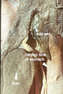
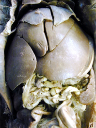
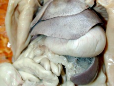
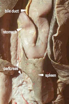
The liver is composed of three lobes, and a greenish gall bladder can be found on the central lobe. These lobes extend posteriorly from the transverse septum. Note the bile duct, from the gall bladder, which goes to the duodenum along with two hepatic vessels. The pancreas, consists of two lobes; a ventral lobe overlying the duodenum, and a dorsal lobe, in the curve between pyloric stomach and duodenum. The pancreatic duct is usually difficult to locate, it runs from the junction of the lobes into the duodenum. The spleen, an organ of the circulatory system (lymphoid tissue), extends posteriorly from the curvature of the stomach.
Dogfish Urogenital System
Excretory System
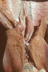
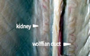
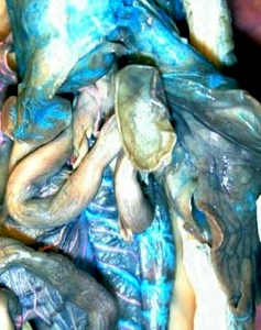
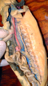
Locate the opisthonephric kidneys on the dorsal body wall. They are flat grey bands running the full length of the body cavity behind all of the other organs and the peritoneum. They are therefore retroperitoneal. The kidneys developed from a strip of nephrogenic tissue derived from mesomere mesoderm. Sharks retain urea, which makes them hyperosmotic. Water flows in from the environment. Rectal (salt) glands secrete excessive salts to maintain osmotic balance. The kidney ducts enter a urogenital chamber, which is partly separated by the urogenital papillae from the rectum in the cloaca.
Genital System
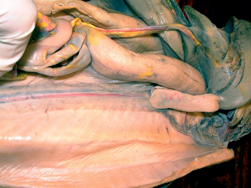
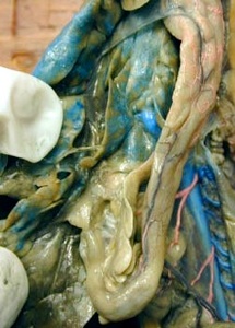
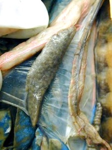
In the male, locate the two large testes on the dorsal body wall, which look like small lobes of the liver. They lead by a series of small ducts called vasa efferentia (which are undeveloped kidney tubules) into the opisthonephric (mesonephric) or Wolffian duct, which is convoluted and on top of the kidney. The urine also goes through this duct. When the duct widens and straightens into a seminal vesicle, the ‘ureter’ forms a bypass for the urine. The sperm sac, which may be full or empty depending on the season, stores mature sperm. These sacs are remnants of Muellerian ducts. The male uses the claspers for internal fertilization. Inside the claspers is a siphon sac that adds sugars to the sperm. Male dogfish mature at about 11 years of age.
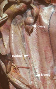
In the female, which matures at 18-21 years, locate the two large ovaries, which look like small lobes of the liver. They may be swollen with eggs covered in yolk. The eggs move from the ovary into the body cavity and then into the opening (ostia) of the two oviducts, called Muellerian ducts. They are silvery tubes on top of the kidneys. The oviductual gland provides albumen and makes egg cases in some species. It may be large or small depending on the season. 6-7 embryos grow in the oviduct (uterus) for 20-24 months being nourished by the yolk, which is absorbed when they are born. They obtain some nutrients and gas exchange from the mother.
NECTURUS
Necturus digestive tract
Use your scissors to make a longitudinal cut off center from the cloaca to the transverse septum. Do not lift as you are trying to keep the mid ventral mesentary intact.
Note the long, mid-ventral falciform ligament connecting the ventral body wall and the liver. Show this to your group before breaking it when you open up the cavity. Cut across the posterior edge of the transverse septum avoiding the pericardial cavity. Like the dogfish, the main digestive tract is in the pleuroperitoneal cavity. The greater omentum extends from the dorsal wall to the stomach and the gastrosplenic ligament goes from the stomach to the spleen. The dorsal mesentery holds the intestines to the dorsal wall; it is called the mesocolon at the large intestine. Other ligaments support the lungs and urogenital tract. Be careful not to destroy these delicate membranes when observing the organs.
Use your scissors to cut through the corner of the left jaw. Cut down just along the ventral side of the gills all the way to the arm. Then join up with the body cavity below the septum. Open up the mouth. Be sure to avoid the central pericardial area and any blood vessels.
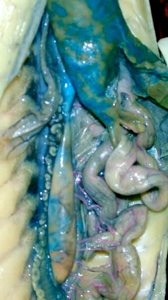
In the oral cavity are teeth, two rows in the upper jaw, and one in the lower. The tongue is better developed than that of the shark. The pharynx has two pairs of gill slits. On the posterior floor of the pharynx lies the glottis, a slit that leads into the trachea. From the pharynx to the stomach is a short, poorly defined esophagus. The straight stomach has internal rugae and ends with a muscular pyloric sphincter. The small intestine is the duodenum anteriorly, and then a long coiled portion with internal small folds called plicae, which increase the surface area. There is a short large intestine, which enters the cloaca via the anus.
Necturus digestive organs
The large liver is not lobed but weakly scalloped posteriorly. The gall bladder is on the underside of the liver near the duodenum. The hepatic ducts and bile duct empties into the duodenum as in the shark, but may be difficult to see. The irregular cream mass of the pancreas lies on the hepato-duodenal ligament and is fused into one. Two small ducts lead to the duodenum. The pancreas is both an exocrine gland, producing digestive enzymes and an endocrine gland regulating metabolism. The spleen, a lymphatic organ, lies to the left of the stomach.
Necturus Urogenital System
Excretory System
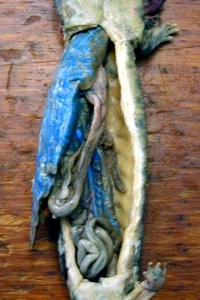
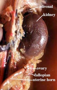
The mesonephric kidneys are long flat strips, narrower anteriorly, which lie dorsally close to the midline, in the pleuroperitoneal cavity. Unlike other vertebrate kidneys, they are not retroperitoneal, but are supported by a peritoneal fold. Urine is produced and collected by tubules, which enter the mesonephric duct. In males this duct is large and coiled in the anterior half as it is used by sperm as well. In females, it is small, on the outside edge of the kidney and separate from the oviduct, but stuck to it.
The bladder is first found in amphibians. It forms as a pocket in the floor of the cloaca. The ureters (mesonephric ducts) open into the cloaca and urine backs up into the bladder when the vent is closed.
Reproductive System
In males, the Muellerian ducts do not mature, but persist as rudimentary (vestigeal) structures. It can be seen anterior to the kidney as a thin, black pigmented duct close to the midline. The testis is attached to the dorsal wall by a peritoneal sheet, the mesorchium. Sperm passes through the upper (genital) portion of the kidney by vasa efferentia ducts, which can only be seen when filled with white sperm. The tubules are collected in a longitudinal duct and by more tubules to the mesonephric duct which joins the cloaca. The papillate cloacal gland secretes gelatinous envelopes, which form around clumps of sperm and make spermatophores for sperm transfer in water. The male deposits these spermatophores in front of a female, who picks them up with her cloacal lips.
In females, the ovaries are folded, thin walled sacs, containing many large eggs. Eggs rupture into the coelom and are wafted by cilia to the large ostia (openings) of the oviduct. The linings of the oviducts have glands, which produce jelly to coat the eggs. The convoluted oviducts are attached to the kidney and body wall by a peritoneal fold, the mesovarium. Eggs are accumulated in the terminal end, called ovosacs until they are laid. The oviducts enter the cloaca.
Spermatophores from the male are collected in the fall and stored in the cloaca until spring when they fertilize the eggs as they are laid. Tadpole larva hatch from the eggs. Mudpuppies are neotenic and retain gills and some other larval characteristics as adults.
Mammals
Mammalian digestive tract
In mammals, the coelomic cavity is divided into thoracic and abdominal cavities by the diaphragm. The thoracic cavity will be examined later. For now do not open the thoracic cavity. The esophagus will be seen later.
Try not to tear any of the mesenteries when doing your dissection
Make an incision in the midventral line through the abdominal wall muscles of the cat or rat from the sternum to about 2 cm anterior to the clitoris or penis. Make lateral incisions at the extremities of this first incision and deflect the flaps.
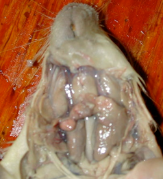
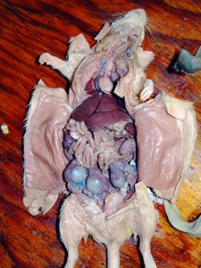
Exposed is the abdominal or visceral cavity. Between the diaphragm and the liver are three ligaments (mesenteries), the most prominent of which is the falciform ligament, which connects the right and left liver halves to the abdominal wall. The greater omentum is double layered and filled with fat in cats. It extends from the greater curvature of the stomach to the dorsal body wall and extends to the pelvic region covering and tucking under the intestine. The portion of this mesentery from the stomach to the spleen is the gastrosplenic ligament. From the lesser curvature of the stomach and duodenum to the liver is the lesser omentum. The dorsal mesentery holds the intestines to the dorsal body wall. Other mesenteries hold the urogenital system in place.
Glands
During the course of your dissection you will remove or view the following glands. For today’s lab, find the three salivary glands, the submaxillary, sublingual and parotid glands, which are located in the head and neck region and are already almost completely exposed. The other glands are not part of the digestive tract, but take the opportunity to observe these neck glands before you open the oral cavity and displace them.
Table of glands and their function
Gland
Location
Function
Submaxillary
central throat, large
salivary
Sublingual
anterior on submaxillary, whitish
mucous
Parotid
towards ear, outside submaxillary, soft
salivary
Lacrimal
below ear
tears
Lymph nodes
4, above submaxillary, dark, round. Others around body.
lymphocytes
Thymus
above heart, large on young animals
lymphatic-disease, immunity
Thyroid
side of larynx, dark red
ion and calcium binding
Adrenal
above kidney
adrenalin or epinephrines
Tonsil
on palate at eustachian tube opening
lymphocytes
Mammalian digestive system
The digestive tract starts with the mouth and its associated salivary glands. Do the glands on the previous page before cutting the jaw.
To view the inside of the mouth, cut through the left jaw after prying the mouth open slightly with a folded paper towel over the teeth. Be sure you are not trying to cut through the molars. To open the lower jaw, cut through the soft palate. Do not open the thoracic cavity.
In the oral cavity note the differentiated teeth and the mobile tongue. Lift the tongue to see the lingual frenulum. Note the papillae on the surface of the tongue. Feel the roof of the mouth with its anterior hard palate and posterior soft
palate. Note the openings to the eustachian tubes and at their opening, the small, soft projection of the palatine tonsils, which produce lymphocytes. The pharynx extends from the oral cavity to the larynx and allows passage of food and air.
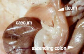
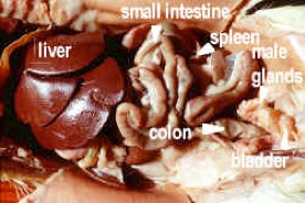
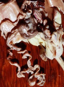
The esophagus ends at the cardiac sphincter and opens into the J-shaped stomach. The pyloric sphincter is at the end of the stomach. The convex side of the stomach is the greater curvature, the concave side is the lesser curvature. The small intestine starts at the pyloric sphincter with the duodenum, a long curved piece, then begins looping back and forth, still descending as the jejunum. The ascending, looping portion is the ileum and it ends in a T-junction with the large intestine. The blind end of the T is the caecum or appendix, large in the rat, small in cats and humans. The other end is the ascending portion of the large intestine or colon followed by a short transverse portion and a descending portion, which ends in a muscular rectum hidden under the pelvic girdle. It opens at the anus, controlled by sphincter muscles, at the base of the tail. The enlarged and constricted areas of the colon are due to peristalsis.
Mammalian digestive organs
The dark reddish brown lobes of the liver (five in rat, six in cat) are attached to the diaphragm. The central lobe has a gall bladder in a cat, but it is absent in the rat, although both animals have a bile duct leading to the anterior duodenum. The mesentery called the greater omentum, stretching from the spleen to the duodenum contains the two lobes of the pancreas, which look like pink granular bubble gum. One lobe runs from the pyloric sphincter to the spleen, the other along the edge of the duodenum. They meet anteriorly forming one duct, which enters the duodenum beside or with the bile duct. The dark reddish brown spleen is an elongate lobe on the left side just below the stomach.
Mammal urogenital system
Excretory System
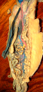
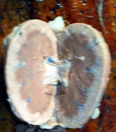
The structures of the excretory system are paired kidneys and ureters from mesomere mesoderm, and an unpaired urinary bladder and urethra. Locate the kidneys on the dorsal part of the abdomen on either side of the vertebral column, embedded in fat. Note a small adrenal gland anterior to each kidney. This is an endocrine gland and is not part of the excretory system. The kidneys have a duct or tube, the ureters leading from them. The kidneys are bean shaped and are metanephric kidneys, which are a new formation. They are retroperitoneal (outside the coelomic space) and came from a strip of mesomere, which formed the caudal end of nephrogenic mesoderm. This piece becomes displaced and moves laterally and anteriorly. The notch or hilus of the kidney is the site of entrance and exit of the blood vessels and the ureters. Section a kidney (like in the diagram) and identify the cortex (outer layer), medulla (inner layer), the pelvis (a cavity) and the papilla of the calyxes, which project into the pelvis. The rat has only one papilla. There are also some models to show these parts. Excretory products in the urine pass into the pelvis, thence to the ureter. Urine may either be hyperosmotic (higher solute, excess salts) to the blood or hyposmotic (higher water). The ureters pass posteriorly and enter the neck of the urinary bladder. The urinary bladder is a pear-shaped sac supported by ligaments (remnants of the ventral mesentery), with a narrowing neck, which terminates in a tube, the urethra that opens to the outside. It is very small in animals like rats which do not hold urine for long periods. In mammals, the bladder comes from the stalk of the allantois (evagination of the embryonic cloaca) and is endodermal in origin. Hormones may allow water reabsorption from the bladder.
Reproductive System
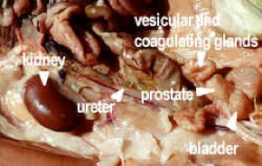
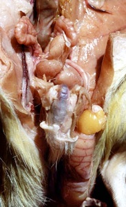
The male reproductive system consists of testes, ducts, glands, urethra and penis. The testes (from hypomere mesoderm) are located in an external pouch, the scrotum, which is a projection from the peritoneal cavity. Scrotal sacs have descended outside the abdominal cavity to keep the sperm cool. Remove this sac on one testis and also the thin, inner peritoneal sac. An epididymis lies on the dorsal surface of each testis, consisting of the head or caput epididymis (which receives sperm from the seminiferous tubules of the testis proper via the vasa efferentia or efferent ductules), the body of the epididymis (which matures sperm) and the caudal portion (which holds sperm like the seminal vesicles of dogfish). The epididymis joins a convoluted tube, the vas deferens. The vas deferens from each testis carries sperm to the urethra. It is looped over the ureters in the region of the neck of the urinary bladder, then turns posteriorly, and enters the urethra a short distance posterior to the bladder.
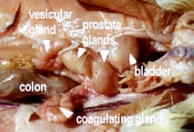
Identify the prostate glands, on either side of the bladder, at the point of entrance of the vas deferens, and Cowper’s glands (bulbo-urethral glands) at the base of the penis. Cowper's glands are buried deep between the posterior and lateral muscles at the base of the penis. The prostate and Cowper’s glands both contribute seminal fluid to the urethra. In the male rat, there are a number of additional prostatic glands. Use the diagram to identify the vesicular, ampullary and coagulating glands.
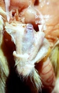
Trace the urethra posteriorly, and identify the penis, glans penis (acorn shaped tip) and the prepuce (foreskin). Note that the neck of the urinary bladder, in males, continues as the urethra, through the penis to the exterior. The tip of the penis has two elongate preputal glands used for lubrication during sperm transfer. The sperm uses the same passageway as the urine in the urethra, but not at the same time due to an automatic shunt in the duct.
The female reproductive system consists of a pair of ovaries, a pair of fallopian tubes, a pair of uteri and a vagina. The ovaries, from somatic hypomere mesoderm, lay posteriorly to the kidneys, held in position by a mesentery, the mesovarium. From each ovary eggs are shed into the coelom and a fallopian tube with a wide mouth, or ostium receives ova. The tubes coil, and then enter the uterus. The uteri from both sides join in the midline to form a "V’ shaped structure. Each arm of the Y is formed by the cornua, or horns of the uterus, and the base is the body of the uterus. Such a type of uterus is termed a bicornuate uterus. The uterus joins the vagina at the posterior end of the short body of the uterus. The projection of the uterus into the vagina is known as the cervix. The urethra is joined ventrally onto the vagina. The common passageway to the outside housing the separate ducts, urethra and uterus is the vestibule. Cut into it and trace the ducts. The external portion of the vagina is the vulva. At the ventral side of the vestibule near its orifice is a small structure, the clitoris, which is the homologue of the penis of the male. In rats, it encloses the end of the urethra and conducts urine to the outside. At the edges of the vestibule are the labia minora derived from genital folds and the labia majora derived from genital swellings, which are homologues to the scrotal sac of males.
Examine the enlarged uterine horns of a pregnant female. Cut them open to see the fetus in the amniotic sac. The fetus is attached to the uterine wall by the umbilical cord, which contains two arteries and a vein. The umbilical cord ends in a zonary placenta for both cats and rats.