
|
Overview of the Secretory Pathway: The secretory pathway involves the flow of material from the Golgi to the cell surface. There are two different secretory processes, constitutive secretion in which proteins are sorted into vesicles in the Golgi and move directly to the cell surface and fuse with the plasma membrane (exocytosis), resulting in the release of soluble proteins to the exterior, or incorporation of membrane proteins into the plasma membrane. The second pathway, regulated secretion, differs in that the proteins are stored in secretory vesicles that later fuse with the plasma membrane in response to a specific signal. Click to enlarge image. |
Each Golgi stack has two faces, the cis face near the ER and the trans face facing away from the nucleus toward the plasma membrane. The flow of material in the Golgi apparatus is from cis to trans. Transport vesicles from the E R containing membrane and soluble proteins fuse with the cis Golgi network. See Figure 14-24. Proteins then move via vesicles from one cisterna to the next. As the proteins move along they are processed and sorted. Vesicles leave the Golgi for a number of destinations. These include:
How are proteins targeted to these three different fates? The fates of the proteins are thought to be determined by features of the proteins themselves.
Protein aggregates containing proteins destined for regulated secretion are seen in vesicles in the process of budding from the trans Golgi network. The aggregation occurs in response to the acidic conditions of the trans Golgi network. Proteins that are normally secreted via the regulated secretory pathway form aggregated in vitro when incubated under the ionic and pH conditions that occur in the trans Golgi network (pH 6.5, 1 mM Ca++). Aggregation does not occur under the pH neutral conditions of the earlier parts of the secretory pathway. These protein aggregates give rise to the 'dense core secretory granules' that are the typical storage vesicles of the regulated secretory pathway.
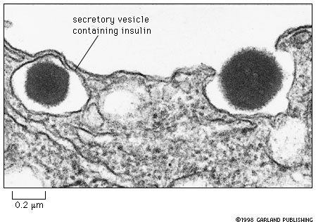 |
Fig. 14-26. Insulin containing dense core secretory in a islet beta cell of the pancreas. One of the granules in being exocytosed. |
The regulated secretory pathway is used for proteins that are stored and secreted on demand. For example, insulin is produced by the beta cells of the pancreas and stored in dense core secretory granules. When blood sugar increases to a threshold level, insulin containing secretory granules fuse with the plasma membrane, releasing insulin to the blood, as shown above. The digestive enzymes produced by the acinar cells of the pancreas are stored in dense core granules called zymogen granules and are released into the duct leading to the gut following feeding.
As noted in the previous lecture on protein processing, proteolytic clevage of many secreted proteins occurs during the maturation of dense core secretory granules.
Dense core secretory granules dock at the cell surface and release their contents when they are stimulated to do so. This usually occurs through the dramatic rise in intracellular Ca++ concentration that occurs by means of a calcium action potential. For example in nerve endings synaptic vesicles containing neurotransmitters (e.g. acetylcholine) fuse with the plasma membrane and release their contents when voltage activated calcium channels open in response to depolarization of the membrane. Calcium action potentials, unlike sodium potentials, are graded, rather than all-or none, and may be quite localized. The sequence of molecular processes behind regulated exocytosis are not fully understood.
A spectacular example of exocytosis of dense core secretory granules that many
students know about from first year biology is discharge of trichocysts in Paramecium.
Trichocysts are compact elongated granules that produce a long stiff thread-like
product on exocytosis. When the cell is strongly stimulated by mechanical or
chemical means, trichocysts discharge. This reaction occurs very quickly. In
a few milliseconds as many as several thousand trichocysts may be discharged.
This synchronous discharge of trichocysts involves a sudden rise in intracellular
Ca++ as Ca++ channels in the plasma membrane and ER membranes
open.
| Trichocyt discharge in Paramecium | |
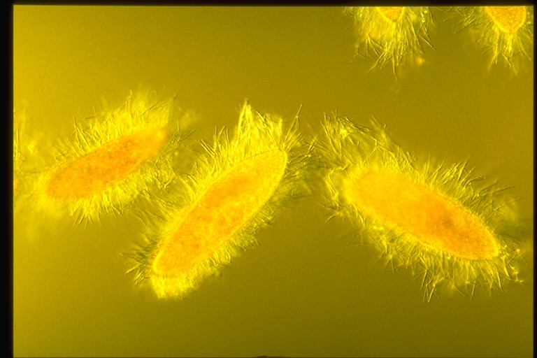 |
These cells have been killed with picric acid. Before they die they discharge trichocysts. This rapid mass exocytosis of dense core secretory granules occurs in a small fraction of a second. Photo by Dr. Yuuji Tsukii, Hosei University. Click to enlarge. |
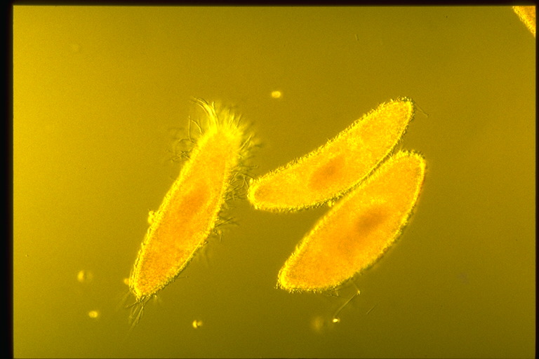 |
This is a control with cells carrying a mutation that blocks trichocyst discharge. notice that there is some discharge in the cell at the left. The mutation is recessive and there are a few wild-type trichocysts that have been carried over from a parent cell. Photo by Dr. Yuuji Tsukii, Hosei University, Click to enlarge |
The constitutive exocytosis pathway operates continually in all cells and supplies a continuous stream of vesicles containing lipids and proteins for the plasma membrane. Other proteins and glycoproteins form part of the extracellular matrix. These materials are secreted via the constitutive secretory pathway. You might read about collagen in animal cells on pp. 600-2.
This is an important part of the material dealing with methods of studying processing of proteins and vesicle transport. You are responsible for this material.
The last ten pages of the chapter (pp. 467-77) give examples of operation of the secretory pathway. You are to study these on your own.
Our expectation is that you will learn the processes in detail using at least one of the following examples:
You should look at this essay question from a previous exam. Outline your own answer before looking at the set of student answers. There is much to be gained from analysis of these answers.
 |
Overview of the Lysosomal Pathway: Lysosomes are vesicles that contain digestive enzymes. These vesicles serve as containers in which digestion of proteins and other macromolecules occurs. The digestive enzymes move through Golgi to Late endosomes (red arrow), In late endosomes proteins are sorted and the receptors are returned to the Golgi (blue arrow) the lysosomal proteins are passed on the to the lysosomes (red arrows). Lysosomes also receive input of proteins from the cell surface (endocytotic pathway) that will be described below. Click to enlarge image. |
The lysosomal pathway directs proteins to lysosomes. Lysosomes are vesicles that contain digestive enzymes and that are the site of intracellular digestion. Their activity will be discussed below in the lecture on intracellular digestion. Proteins destined for lysosomes have a special targeting signal. They are all glycosylated and some of the mannose residues in the attached oligosaccharides are phosphorylated to form mannose-6-phosphate. Mannose-6-phosphate is the targeting signal for lysosomal proteins. The trans Golgi network contains a special mannose-6-phosphate receptor that binds to and results in the concentration of proteins carrying oligosaccharides bearing mannose-6-phosphate into vesicles that are destined for lysosomes. The mannose-6-phosphate receptors are recycled in an interesting process that will be discussed below.