Chapters
Lodish 4th Edition: Chapter 21 pages 917 - 921
Moyes and Schulte: Chapter 5 pages 146 - 164 plus review Chapter 3 pages 87-89
Introduction to nerve signaling
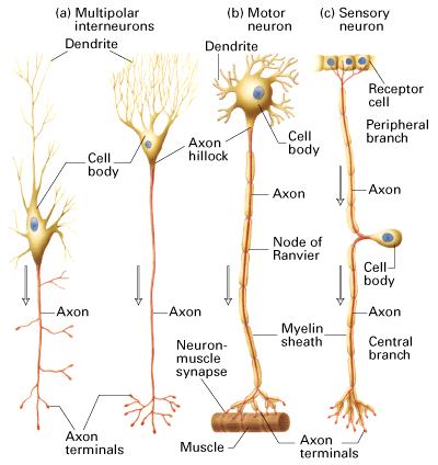
Figure 21-1. Lodish 4th edition

Figure 7-29. Lodish 5th edition
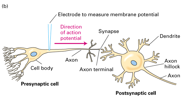
Figure 21-2. Lodish 4th edition.
Neurons come in different shapes and sizes. They generate can generate action potentials and/or passive potentials. In addition neurons communicate with other neurons or cells (such as muscle) with synaptic connections. These can be either chemical or electrical synapses.
Passive membrane potentials: neuronal perspective
All cells have a membrane potential (an electrical potential) that exists
across the cell membrane
To understand how synaptic or nerve communication is generated and modified
you must understand how resting membrane potential is established and
maintained
Researchers use microelectrodes to measure the voltage difference between the
outside and inside of the cell
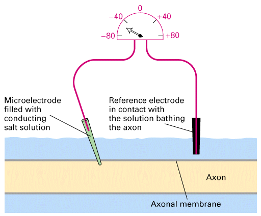
Figure 21-7, Lodish 4th Edition OR Figure 7-14, Lodish 5th edition.
A microelectrode, constructed by filling a glass tube of extremely small diameter with a conducting fluid such as KCl, is inserted into an axon in such a way that the surface membrane seals itself around the electrode. A reference electrode is placed in the bathing medium. A potentiometer connecting the two electrodes registers the potential. The potential difference maintained across the cell membrane in the absence of stimulation is called the resting potential, in this case, -60 mV. A potential difference is registered only when the microelectrode is inserted into the axon; no potential is registered if the microelectrode is in the bathing fluid.
Nernst equation: is used to calculate the exact electrical potential
that is generated for a known concentration difference in a specific ion,
separated by a membrane permeable to that ion.
Ex= RT ln [X]o
.......zF.....[X]i
OR at room temperature (22oC)
Ex= 58 log10 [X]o
......................[X]i
can measure the membrane potential of a cell = the voltage difference
between the inside and the outside of the cell.
The resting membrane potential is set by a combination of a number of
different ions.
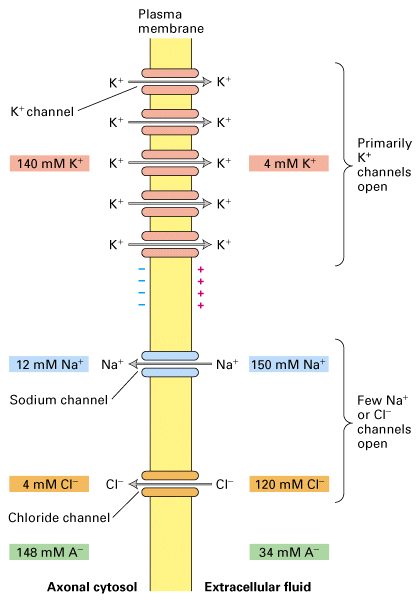
Figure 21-9, Lodish 4th edition. Origin of the resting potential in a typical vertebrate neuron.
The ionic compositions of the cytosol and of the surrounding extracellular fluid are different. A- represents negatively charged proteins, which neutralize the excess positive charges contributed by Na+ and K+ ions. In the resting neuron there are about ten times more open K+ channels than open Na+ or Cl- channels; as a consequence more positively charged K+ ions exit the cell than Na+ or Cl- ions enter, and the outside of the plasma membrane acquires a net positive charge relative to the inside.
K+ is dominant because the permeability to K+ (PK) is the
greatest. This is because there are leak K+ channels found in the membrane (of
all animal cells) that allow the passage of K+ across the hydrophobic lipid
bilayer. Therefore the resting membrane potential is closest to the Nernst
potential for K+.
But resting potential is not identical to EK and therefore other
ions must have an influence.
There is actually some leakage through Na+ and Cl- channels too. Therefore
there is some permeability to Na+ (PNa) and Cl- (PCl-).
Actual measurements of membrane potential
Experimenters tested the theories out by measuring resting membrane potential of giant axon of squid by placing recording electrodes into the axon.
They gound that Vm (at r.t.) = -65 to -70 mV (close to the potential calculated for K+ ions but not exactly at EK+)
Therefore they decided to test the influence of external potassium
concentration on resting membrane potential by increasing [K+]external and
measuring the new membrane potential
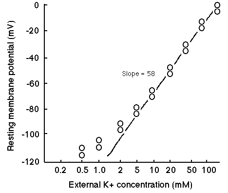
graph: slope = -58 mV
- this means for every ten fold increase in external K+ concentration the potential increases by 58 mV at r.t.
- graph is pretty close to theoretical numbers except at lower external K+ concentrations (which is what is found in nature)
- another ion is influencing the resting membrane potential
- the membrane is also permeable to Na+ ions (also Cl- but has much less influence on resting membrane potential)
- the membrane permeability is much lower for Na+ ions than K+ ions
Permeability = presence of channels that allow ions to flow in or out of cell "leak channels" or "rest channels"
- could just be common proteins found in all cells that allow ions to flow or specialized proteins that function to create nerve membrane permeability
- Na+ ions flow into cell down concentration gradient
- cause a slight depolarization of membrane away from K+ equilibrium
- therefore Vm of squid axon is -65 mV instead of -93 mV
- currents carried by inward leak of Na+ and outward leak of K+ are balanced at equilibrium and the resting potential is stable
Calculation of resting membrane potential
The membrane potential can be considered a combination of all three ions
such that:
The Goldman-Katz equation can combine the Nernst equations for all three:
Vm = 58 log10 PK[K+]o + PNa[Na+]o + PCl[Cl-]i
........................PK[K+]i + PNa[Na+]i +
PCl[Cl-]o
OR
Vm = 58 log10 [K+]o + [Na+]o(PNa/PK) + [Cl-]i(PCl/PK)
........................[K+]i + [Na+]i(PNa/PK) +
[Cl-]o(PCl/PK)
In the squid axon at rest: PK : PNa : PCl = 1 : 0.04 : 0.45
Therefore the membrane potential can dramatically change if the permeabilities
to the ion changes.
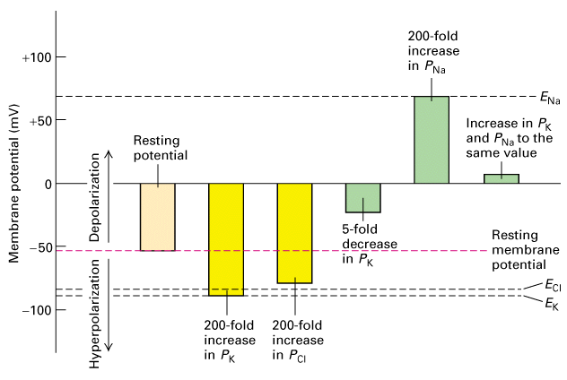
Figure 21-10. Lodish 4th edition. Effect of changes in ion permeability on membrane potential.
The resting membrane potential is -53 mV; ENa, EK, and ECl are the potentials calculated from the Nernst equation if the membrane contains only open channels for Na+ or K+ or Cl-, respectively.

Another view of Nernst potentials with respect to resting membrane potentials
Passive flow of current
Signals can be transmitted through a neuron either through passive current flow or using an action potential. Passive flow of current can be thought of in terms of a current traveling down a copper wire.
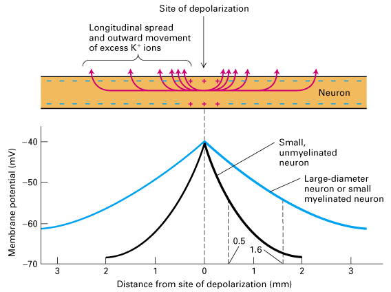
Figure 21-11. Lodish 4th edition. Passive spread of a depolarization of a neuronal plasma membrane with only resting K+ ion channels.