|
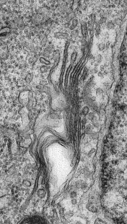
Golgi section in a cell of the accessory boring organ of the
marine snail Ocaenebra. Click on the little image at the left to
bring up a larger figure. |
 |
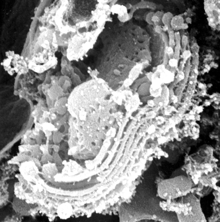
This is a freeze-etch preparation showing golgi and the trans-golgi
network from a cell of the accessory boring organ of the marine snail Ocaenebra.
This picture and that above are both of the same organelle in the same cell
type. What features are shown to better advantage here than in the figure
above? Which are shown better in the section (above). |
 |
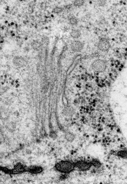
Golgi in Sainouron, a protist. Don't worry about the name.
Look at the Golgi |
 |
This is a golgi (dictyosome) adjacent to the forming cell
wall in a tobacco leaf. |
 |
This is a golgi (dictyosome) in another protist (bodonid). |
 |
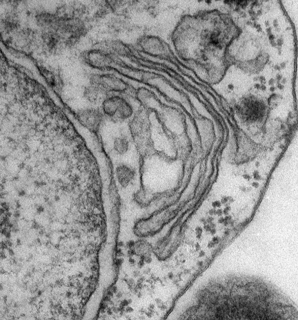
Golgi in rat liver cell |